


 Department Overview
 Doctors
 Technology
 Research
Department Overview
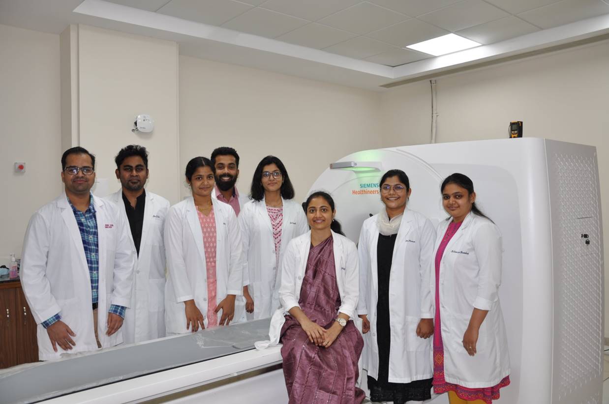
The department is headed by Dr B T Pushpa who with seven additional radiologists and MSK radiology fellows ably manage the high turnover and huge demands of the busy clinical divisions of the hospital.
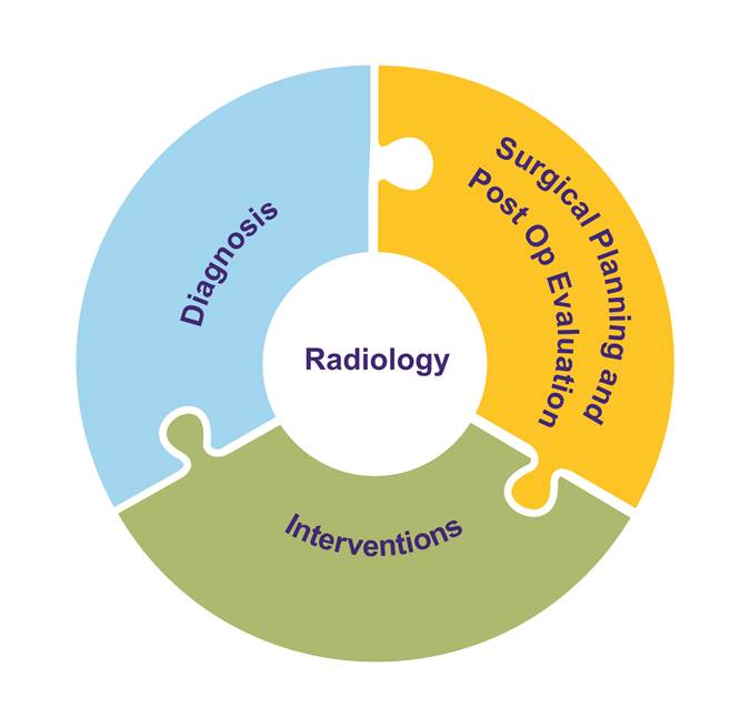
Radiology involves the use of advanced imaging technology that helps in management of patients at all stages of diagnosis, surgical planning, intraoperative guidance and follow up evaluation. With the recent advances in basic science and various modalities of imaging, radiology forms an integral and essential part of day to day practice in Ganga Hospital
The hospital is a tertiary referral centre drawing referrals of complex and challenging cases from all over India and neighbouring countries and the radiology department forms an important hub contributing to the various specialties of imaging of polytrauma and major injuries, and all musculoskeletal disorders including spine, sports medicine, pediatric orthopaedics, joint diseases, hand disorders and oncology.
While upholding the standards of safety and patient-centered care, the department of Radiology & Imaging at Ganga Hospital provides a full spectrum of diagnostic and interventional radiology treatments.
The Department is available 24 hours a day, 7 days a week, and boasts the most cutting-edge, modern facilities in Coimbatore. The radiologists and radiology techs on the team have subspecialty knowledge in addition to general skills
The data below clearly shows the huge turnover in the field of musculoskeletal radiology which can be matched only by a very few other centres in the world.
The department possesses the following equipment which help our experts to cater to a large number of challenging imaging requirements.
The Various Radiology Techniques Performed 2015 - 2022
| Year | 2015 | 2016 | 2017 | 2018 | 2019 | 2020 | 2021 | 2022 |
|---|---|---|---|---|---|---|---|---|
| MRI | 7251 | 8477 | 8613 | 8780 | 8622 | 6994 | 10332 | 11583 |
| CT | 7022 | 7621 | 8266 | 8505 | 8896 | 8025 | 7807 | 9937 |
| USG | 2967 | 7122 | 3869 | 3702 | 5220 | 4977 | 4222 | 5640 |
| X-Ray | 1,18,599 | 1,21,974 | 1,13,530 | 1,12,985 | 1,35,677 | 88,201 | 123181 | 143335 |
| Intervention Procedure | - | - | - | - | 230 | 409 | 884 | 1464 |
| Mammogram | - | - | - | - | 63 | 65 | 126 | 13801 |
Our Doctors


Specialization
- Anesthesiologists As Peri-operative Physician
- On Arrival Nerve Blocks in Trauma
- Regional Anaesthesia
- Anaesthesia for Major Spine Surgery
- Anaesthesia for Joint Replacement
- Anaesthesia for Paediatric Orthopaedics
- Anaesthesia for Hip Fractures
- Poly Trauma Management
Technology
MRI forms an essential part of evaluation of the musculoskeletal system. Having the advantage of no radiation and the ability to clearly define the status of bone and all soft tissues, it is the first choice for complete evaluation of the spine, joints and nervous system.
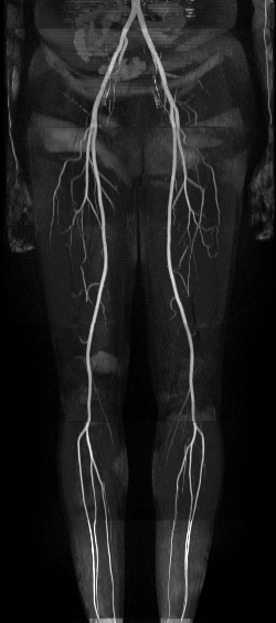
Non Contrast MR Angiogram of both Lower Limbs
The 3T Lumina platform acquired by the hospital recently is one of the latest in the world and with a special MOU with SIEMENS, Germany. IT has facilities for advanced sequences like Magnetic Resonance Spectroscopy, T1 rho, T2 relaxometry and UTE which are capable of not only looking at tissues with greater precision but are also capable looking at molecular variations taking radiological evaluation to the next generation of imaging. The systems are also loaded with special programs, Best examples are angiogram of the lower limbs , renal arteries without use of contrast. Its wide bore design is well accepted by the claustrophobic patients. There are plenty of advancements available with machine which adds to the safely of the patients by changing the way it scans depending on the patients biometry.
Two CT scans (Seimens Emotions 6, Somatom Go Now) which are employed for the day to day evaluation of patients with polytrauma and major injuries and complex orthopaedic and spinal problems. The machines are capable of performing high quality 3D images and also angiograms to evaluate vascular integrity.
Non Contrast MR Angiogram of both Lower Limbs
Our radiologists and technicians are also committed to your safety and have evolved numerous clinical and radiological protocols that reduce radiation exposure to the minimum. The CT machines are also provided with the latest dose reduction softwares that ensure the maximum reduction of radiation to every patient.
Apart from routine USGs, department is well focused on joint and soft tissue ultrasound avoiding unnecessary MRI or CT scan.
The department caters to more than 700 patients who require plain radiographs every day. As each patient may require a few to many X-ray exposures, the number of the Xray exposures per day frequently crosses 2000 per day. This requires machines which can handle this high turnover and also provide high quality digital images which are available without any delay and with high clarity. To meet this demand, the department has 12 state-of-the-art digital X-ray machines which include three ceiling mounted systems that allow special image acquisitions and stitching facility which are required for ‘Standing whole spine X-ray, whole limb alignment radiographs and specialized views of the limbs in major trauma.
Mammography is X-ray imaging of the breasts. For Further details Click here
Various image guided procedures are performed in department for diagnostic as well as therapeutic purpose.
Diagnostic procedures : Image guided biopsy and needle aspirations are accurate and safe since it is performed under image guidance.
Therapeutic procedures : Therapeutic procedures give quick results and are least invasive as compared to surgery. Most patients benefit from these procedures as they obtain rapid relief of pain which help in continuing physiotherapy and to avoid oral medications.
USG guided procedure :
Joint injections : These are particularity help help in age related changes in the joint and to avoid surgery as much as possible. People with Stiff shoulder have rapid and good relief by injection of medications directly into the joint. Tendon injections: Tennis elbow, golfers elbow, Achilles tendon problems, and plantar fasciitis can be effectively treated by targeted injection of the medications through ultrasound guidance.
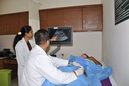
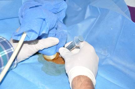
USG Guided Hip Injection
Nerve root injections : Most accepted and well received by the patient is ultrasound guided nerve root injections in cervical disc prolapse. This might avoid surgery in many patients and give rapid pain relief.
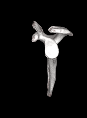
CT guided procedures : Problem associated with backache like degenerative changes in sacroiliac joints, facet joints, pain in the tail bone regions are treated by CT guided selective joint and nerve root injections. Complex and difficult to access areas in body can be approached by CT scan guidance for biopsy and drainage procedures.
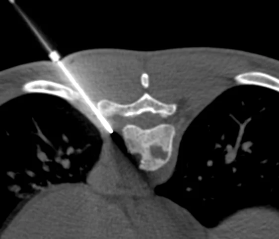
CT Guided Biopsy
- Siemens Lumina 3T MRI
- Siemens Magnetom Symphony 1.5 Tesla MRII
- Siemens MDCT (Somatom 128 slice)
- Siemens Magnetom Symphony 1.5 Tesla MRISiemens Multi-Slice CT (Somatom Emotion)
- Esoate (Mylab 5)
- Philips Affinity 70
- Siemens Acuson 500
- Wipro GE Logiq S7
- Siemens Digital X-Ray
- Digital X-Ray
- Siemens Mobile X-Ray
Wipro GE DEXA Scan OPG (Myray) Hyperion Hologic 3D Digital Mammogram
- AIRO - intra-operative CT navigation
- Siemens
- GE
- Sonosite
- Mindray
- Siemens Acuson 500
- Wipro GE Logiq S7