Department Overview
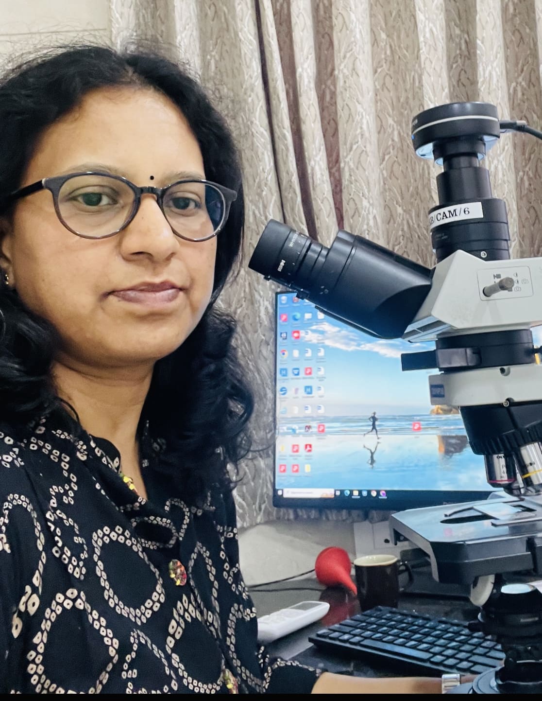
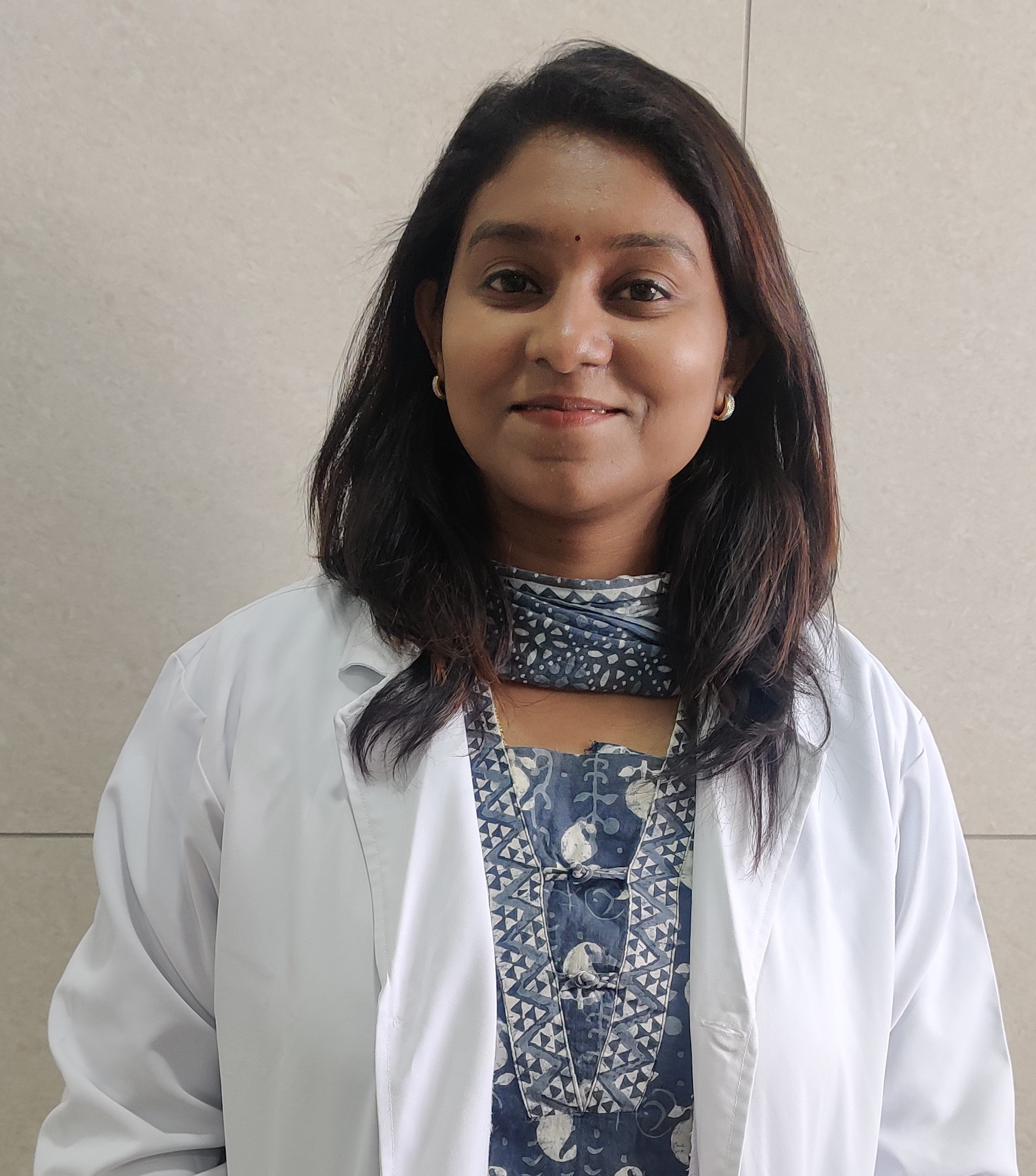
Our Ganga Hospital is proud to have an excellent in-house pathology department headed by Dr Sindhu R , who is specialised in cancer pathology. The great advantage of having an in-house pathology department is that it allows faster processing time, better communication between the pathologist and the oncologists, facilitates clearer and quicker decisions
Pathology is a medical speciality that deals with the diagnosis of disease. Although most illnesses are diagnosed clinically and radiologically, diseases, especially cancer, are confirmed by pathological examination. This is done by macroscopic, microscopic, immunologic and molecular analysis of organs, tissues and body fluids.
Physical and radiological examination often provide only part of the picture of an illness. Pathology helps to confirm the diagnosis. It also lets us know the subtype of cancer and the various properties of the cancer tissue. This greatly helps us to plan our treatment. For example, when we send a tissue of breast cancer for biopsy, the pathology helps us confirm that it is cancer and know the type of cancer, the aggressiveness of cancer, and whether it has hormone receptors. This allows us to plan the course of treatment for the patient. In addition, pathology helps us know whether cancer has been fully removed or not and assess the margins removed for each tumour.
Histopathology including Immunohistochemistry
Cytopathology
Hematopathology
Intraoperative consultation (Frozen section ).
Histopathology is the branch of surgical pathology that involves diagnosing a disease by examining the sampled tissues under the microscope by using a glass slide. This helps us to confirm cancer and understand the behaviour of the cancer cells.
It is a quick, inexpensive and straightforward procedure wherein a needle is inserted into a superficial lump or swelling. A small number of cells are acquired by negative pressure using a syringe. These cells are examined under the microscope to determine whether the lump is benign or malignant(cancer). If the mass is deeper, the same procedure can be done under ultrasound guidance. This is an office procedure and can be done in the outpatient clinic. It causes minimal pain and carries virtually no risk of complications.
The standard processing of tissues for pathology takes about 48 hours. However, while operating, we would like to know the confirmatory diagnosis while doing the operation as this would determine the operation that we would do. Therefore, the frozen section freezes the tissue of interest, helps swiftly process the tissue and give the report early during the ongoing surgery so that the surgeons can continue with the operation. For example, while doing breast cancer surgery, if we come across a slightly enlarged lymph node, it could either be due to cancer or low-grade infection. If it is due to cancer, then the large axillary tissue needs to be removed. If it is due to a low-grade infection, no further surgery is required. In these instances, the frozen section helps tell the surgeon immediately whether the involved lymph node is due to cancer or low-grade infection, and the appropriate surgery can be done.
Immunohistochemistry helps us identify specific proteins(antigens) on cells to determine the type of cancer.
Example: In breast cancer, it helps us identify the presence of oestrogen and progesterone receptors. When these receptors are present, it allows us to initiate hormone therapy for these patients


 Department Overview
 Doctors
